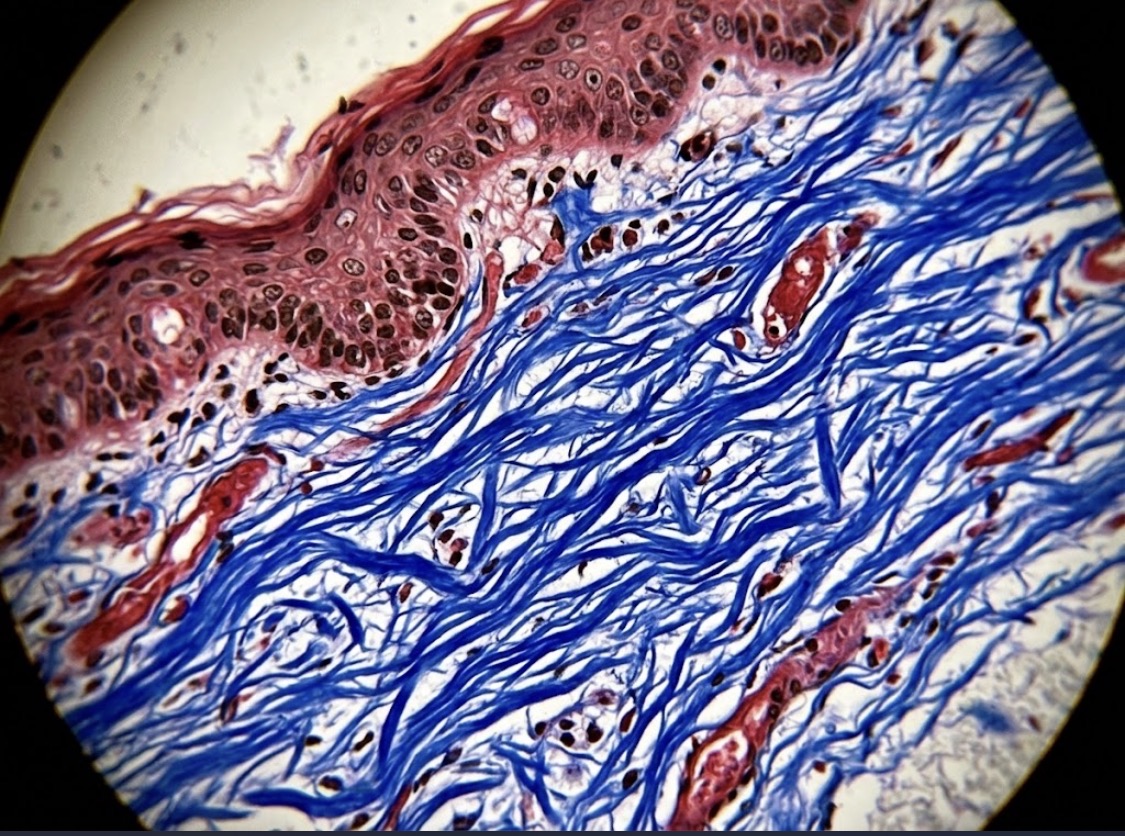
Examine this tissue section. What staining technique was used to highlight collagen fibers in blue?
LabInview
13 days ago•19 Total Votes· Final Results•2 Responses
Select Your Diagnostic Answer
Ended · Final Results
Clinical Discussion & Responses (2)
Responses from the community about this case.
Sign in to join the clinical discussion.
Victoria Folorunsho
13 days ago
Masson's Trichrome stain collagen fibres blue, while the muscle and cytoplasm stain red and the nuclei appear dark purple or black
Haruna Ali
13 days ago
- *Masson's Trichrome:*
- *Collagen / Connective tissue = Blue or Green* (depending on variant)
- *Cytoplasm / Muscle / Keratin = Red*
- *Nuclei = Black / Dark brown*
*Other stains that give Blue Collagen:*
- *Mallory's Trichrome:* Collagen = Deep Blue
- *Aniline Blue* (part of Masson's)
- *Gomori's Trichrome:* Collagen = Blue/Green
- *Martius Scarlet Blue (MSB):* Collagen = Blue
So if you see *Blue collagen in your Histology slide*, answer is almost always:


Clinical Discussion & Responses (2)
Responses from the community about this case.
Sign in to join the clinical discussion.
Masson's Trichrome stain collagen fibres blue, while the muscle and cytoplasm stain red and the nuclei appear dark purple or black
- *Masson's Trichrome:* - *Collagen / Connective tissue = Blue or Green* (depending on variant) - *Cytoplasm / Muscle / Keratin = Red* - *Nuclei = Black / Dark brown* *Other stains that give Blue Collagen:* - *Mallory's Trichrome:* Collagen = Deep Blue - *Aniline Blue* (part of Masson's) - *Gomori's Trichrome:* Collagen = Blue/Green - *Martius Scarlet Blue (MSB):* Collagen = Blue So if you see *Blue collagen in your Histology slide*, answer is almost always: